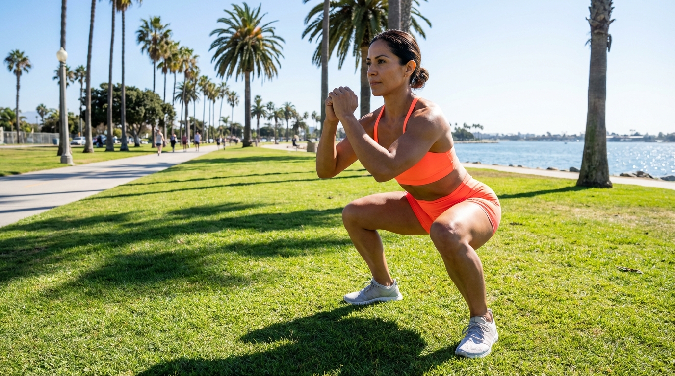
Bodyweight Squat
The fundamental pattern of human movement. A prerequisite for all loaded lower-body training, requiring the integration of triple flexion and kinetic chain stability.
Biomechanics Analysis
Kinetic Chain & Joint Angles
The squat is a Closed Kinetic Chain (CKC) exercise where the foot is fixed to the ground. The movement is defined by synchronous Triple Flexion during the eccentric phase and Triple Extension during the concentric phase.
- Acetabulofemoral (Hip) Joint: Requires θ ≈ 100°–120° of flexion. The posterior displacement of the hips engages the gluteal complex.
- Tibiofemoral (Knee) Joint: Requires θ ≈ 110°–130° of flexion. Knee translation allows the hips to descend while maintaining the Center of Pressure (CoP) over the mid-foot.
- Talocrural (Ankle) Joint: Critical requirement of θ ≈ 20° dorsiflexion. Limited mobility here often forces compensatory lumbar flexion.
Forces & Stabilization
Moment Arms (τ = F × d): As the body descends, moment arms act upon the hip and knee joints. In a bodyweight squat, the torso is typically more upright compared to a low-bar back squat, distributing torque relatively evenly between the knee and hip extensors.
Lombard’s Paradox: A unique biomechanical phenomenon occurs where the rectus femoris and hamstrings—antagonistic bi-articular muscles—contract simultaneously to facilitate extension at both the hip and knee.
Center of Pressure (CoP): Equilibrium is maintained only if the vertical projection of the Center of Mass (CoM) falls within the Base of Support (BoS). The CoP must remain balanced over the mid-foot to prevent anterior or posterior loss of balance.
Muscle Activation Map
Primary Movers (Agonists)
-
Quadriceps Femoris Vastus Lateralis, Medialis, Intermedius, and Rectus Femoris drive knee extension.
-
Gluteus Maximus The primary hip extensor, generating maximum force out of the bottom position ("the hole").
Synergists
-
Adductor Magnus Acts as a powerful hip extensor, particularly in deep flexion angles.
-
Soleus Assists in returning the tibia to neutral from dorsiflexion.
Stabilizers
-
Core Complex Transverse Abdominis and Erector Spinae create Intra-Abdominal Pressure (IAP) to support the lumbar spine.
-
Gluteus Medius Prevents valgus collapse by abducting and externally rotating the femur.
Execution Protocol
-
1
Stance Setup
Position feet between hip-width and shoulder-width apart. Rotate toes outward slightly (5°–15°) to accommodate individual hip anatomy and allow the femur to track cleanly into the acetabulum.
-
2
Brace and Align
Stand tall with full hip and knee extension. Inhale deeply into the diaphragm to create Intra-Abdominal Pressure (IAP). Engage the core musculature isometrically.
-
3
The Descent (Eccentric Phase)
Initiate movement by simultaneously unlocking the hips and knees ("breaking at the hips"). Descend under control (2–3 seconds). Ensure the knees track in line with the second and third toes to prevent valgus stress.
-
4
Depth Achievement
Continue descent until the hip crease is aligned with or below the top of the knee (femur parallel to the transverse plane). Maintain a neutral spine; avoid rounding the lumbar (butt wink).
-
5
The Ascent (Concentric Phase)
Drive forcefully through the mid-foot. Extend the hips and knees simultaneously. Keep the chest elevated to maintain the back angle constant until the hips begin to rise.
-
6
Lockout
Return to the starting position with full extension of the hips and knees. Exhale forcefully at the top. Reset bracing before the next repetition.
Common Mistakes & Corrections
| Fault | Biomechanical Consequence | Correction |
|---|---|---|
| Valgus Collapse (Knees Caving In) | Places excessive stress on the Medial Collateral Ligament (MCL) and meniscus. Indicates weak Gluteus Medius or poor motor control. | Cue "spread the floor apart" with your feet. Actively drive knees outward to track over the toes. |
| Heels Rising (Knee Dominance) | Shifts CoP to the toes (metatarsals), increasing shear force on the knee (patellar tendon) and reducing glute activation. Usually due to limited ankle dorsiflexion. | Focus on "tripod foot" pressure (heel, big toe, little toe). Improve ankle mobility or widen stance slightly. |
| Lumbar Flexion ("Butt Wink") | Posterior pelvic tilt at the bottom of the movement. Compromises spinal integrity under load and places shear force on lumbar discs. | Reduce depth to maintain neutral spine. Improve hamstring flexibility and motor control. Ensure core bracing is maintained. |
| Excessive Forward Lean | Increases the moment arm on the lower back (Erector Spinae) while decreasing load on the quadriceps. Often a compensation for weak quads or stiff ankles. | Keep the chest proud. Imagine a string pulling the crown of the head upward. Ensure knees translate forward enough to allow hips to sit down, not just back. |